Retinal Vascular Diseases
Specialized Care

Retinal Vascular Disease Treatment

Advanced management of diabetic retinopathy, retinal detachment, and vein occlusion.

Book Appointment
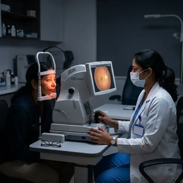
About This Treatment

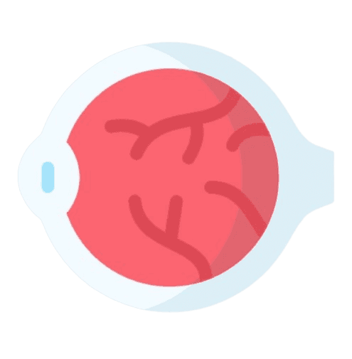
The retina is a thin layer of tissue that contains millions of light-sensitive cells (rods and cones), and is located in the back of the eye. The primary function of the retina is to receive, organize, and send visual information to your brain, through the optic nerve. This whole process enables you to see.

Retinal Disease Impact

Retinal diseases cause damage to any part of the retina. Untreated retinal diseases can lead to severe vision loss and even blindness. With early detection, some retinal diseases can be treated, while others can be controlled or slowed down to preserve, or even restore vision.

Types of retinal diseases:

There are several diseases of the retina. Some of them include:

  • Diabetes-related retinopathy.
  • Retinal detachment.
  • Retinal tears or holes.
  • Retinal vein occlusion, including central retinal vein occlusion and branch retinal vein occlusion.
  • Infectious retinitis.
  • Central serous retinopathy.
  • Birdshot chorioretinopathy and other inflammatory diseases.

There are diseases that affect your macula, such as:

  • Macular pucker, also called an epiretinal membrane.
  • Macular hole.
  • Age-related macular degeneration.
  • Central serous chorioretinopathy
  • Macular edema (swelling due to fluid buildup).

You can inherit other retinal diseases, like:

  • Retinitis pigmentosa.
  • Usher syndrome, an inherited condition that causes retinitis pigmentosa.
  • Stargardt disease.
  • Retinoschisis, which can be something that you’re born with or something that develops over time.

Treatment Options

Treatment depends on the type of retinal disease you have. Depending on the condition, treatment can stop the disease or slow down how quickly it gets worse. Therapy options include surgical procedures, laser treatments and injections of medicines into or around your eyes.

Retinal Vascular Diseases

Book Consultation

Expert care for Retinal Vascular Diseases by our senior specialists.

Schedule Now

Common FAQs

1. Newborn infants are able to see, but as they use their eyes during the first months of life, vision improves. During early childhood years, the visual system changes quickly, and vision continues to develop. If a child cannot use his or her eyes normally, vision does not develop properly and may even decrease. After the first nine years of life, the visual system is usually fully developed and usually cannot be changed. 2. The development of equal vision in both eyes is necessary for normal vision. Many occupations are not open to people who have good vision in one eye only. If the vision in one eye should be lost later in life from an accident or illness, it is essential that the other eye have normal vision. Without normal vision in at least one eye, a person is visually impaired.
Most paediatricians test vision as part of a child’s medical examination. They may refer a child to an ophthalmologist if there is any sign of an eye condition. The American Academy of Ophthalmology and the American Academy of Paediatrics recommend the first vision screening occur in the hospital as part of a newborn baby’s discharge examination.Visual function (including ocular alignment, etc.) also should be checked by the paediatrician or family physician during routine well-child exams (typically at two, four, and six months of age). Later amblyopia and repeat alignment screenings should take place at three years of age and then yearly after school age. If you suspect your child suffers from decreased vision: 1. Amblyopia (poor vision in an otherwise normal appearing eye), refractive error (nearsightedness or farsightedness), or strabismus (misalignment of the eye in any direction). 2. If there are hereditary factors that might predispose your child to eye disease, please make an appointment with our paediatric ophthalmologist as soon as possible. 3. New techniques make it possible to test vision in infants and young children. 4. If there is a family history of misaligned eyes, childhood cataracts, or a serious eye disease, an ophthalmologist can begin checking your child’s vision at a very early age.
Adults should undergo eye examinations at the following intervals: 1. Individuals aged 18-39 with no vision problems or risk factors should have their eyes examined every 1-2 years. 2. Adults over 40 years old with no additional risk factors should schedule annual eye exams. 3. Patients with underlying health conditions, such as diabetes, hypertension, or other systemic diseases, should undergo eye examinations every six months as they are more at risk of ocular complications. 4. Even individuals with glaucoma or a strong family history of glaucoma need prompt check-ups.

Ready to get started?

Schedule your appointment today and take the first step towards better vision.