

Diabetic Retinopathy Treatment in Bangalore
Early screening and treatment to manage vascular damage caused by high blood sugar.
Book Appointment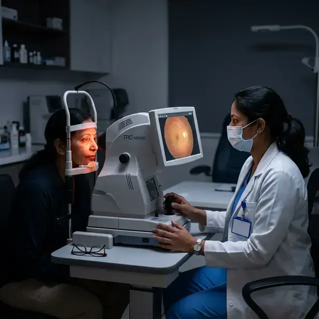
About This Treatment
The blood vessels in the retina are tiny/delicate and prone to damage in patients with uncontrolled high blood sugar levels. The deformation/damage to blood vessels leads to structural changes of the retina termed as Diabetic Retinopathy.
Asymptomatic Nature
A sizeable diabetic population develops Diabetic Retinopathy. The patients initially have no symptoms and have perfect vision even though there would be bleeding and swelling of the retina. If ignored can lead to loss of vision. It is advisable to get retina examined regularly even though the vision is normal.
Symptoms & Causes of Vision loss
- Blurred Vision
- Floaters or dark spots
- Visual impairment
- Difficulty to see at night
- Difficulty in distinguishing colours
Loss of vision is caused by
Diabetic Macular Edema: The weakened blood vessels leak and accumulate fluid in the retina which causes swelling and exudation in the macula of the retina causing loss of vision.
Proliferative Diabetic Retinopathy (PDR): Due to the weakening of the blood vessels, new abnormal blood vessels grow resulting in bleeding in the vitreous. Vitreous is a jelly-like substance in front of the retina. A severe vision loss occurs due to the bleeding in the vitreous.
Treatment may include:-
Timely treatment can stop the progression of the disease. Controlling blood sugar levels can help control the progression of the disease and regular monitoring.
Medical management
Steroid or anti-VEGF Injections into the eye may help reduce swelling of the macula, slowing vision loss and perhaps improving vision.
Laser
If the disease advances, the blood vessels can leak blood and fluid into the retina, leading to macular edema. Laser treatment can stop leakage by targeting specific vessel in the macular or wide spread blood vessel growth(PDR) in Retina.
Surgical management
Vitrectomy involves removing scar tissue and blood from the vitreous fluid of the eye.
Other Services

Cataract Surgery
Advanced surgery to remove clouding lens and restore vision through safe lens replacement.
Micro Incision Cataract Surgery (MICS)
Advanced approach to cataract surgery through incision less than 1.8 mm, leading to improved outcomes.

Phacoemulsification Cataract Surgery
Modern ultrasound-based cataract removal using tiny probes for painless vision restoration.
Book Consultation
Expert care for Diabetic Retinopathy Screening by our senior specialists.
Schedule NowCommon FAQs

Ready to get started?
Schedule your appointment today and take the first step towards better vision.